What is bioluminescence? The fascinating natural glow that illuminates the darkness
A number of living organisms, such as fireflies and jellyfish, produce light as a result of a chemical reaction within their bodies, called bioluminescence.

Food that glows in the dark, fluorescent sheep, or plants that serve as public lighting are things that could well be taken from a science fiction story or even far from reality. But these are not inventions that are the product of someone's imagination, but those that are part of the advances that science has conquered step by step, through years of research on the subject of bioluminescence, which can now give the characteristics of its own light to organisms beyond fireflies and jellyfish.
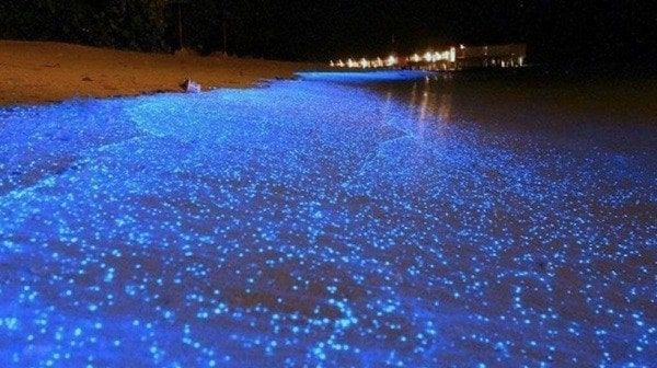
Certain living organisms produce light as a result of a chemical reaction within their bodies, a process called bioluminescence. In bioluminescence, the oxidation of a protein substrate is catalyzed by an enzyme. There are two bioluminescent organisms that have been investigated: fireflies and jellyfish.
The former synthesize the substance called luciferin, which is oxidized with the help of the enzyme luciferase. This reaction is carried out with practically no loss of energy. Jellyfish possess a protein capable of receiving high-energy light (usually in the UV range), called green fluorescent protein (GFP), which emits fluorescence in the green light range. Bioluminescence is quite widespread in marine habitats. More than 90% of animal species in the mid-and abyssal portions of the ocean are thought to emit some form of bioluminescence. On land, it is limited to the fungal and invertebrate kingdom.
Green Fluorescent Protein (GFP)
GFP is produced by the jellyfish Aequorea victoria. The gene encoding this protein, which has already been cloned, is used as a marker in molecular biology. The discovery and study of GFP expand the capabilities of the optical microscope and brings a new visible dimension to the human eye.
GFP can be used as a marker for biomolecules and to follow processes such as cell migration. Thanks to this use, the detrimental effect of chemical fluorescent markers, which were used in research, has been reduced. A fluorophore, after a certain time of exposure to light, releases an electron that reacts with oxygen, originating toxic radicals that would damage the cell and even cause its death. The structure of GFP avoids this impact because when the fluorophore releases an electron, the resulting radicals remain inside the protein without touching the cell.
An important feature of GFP is that it does not need additives to glow, in contrast to other bioluminescent proteins; it is sufficient to irradiate it with UV or blue light for it to emit fluorescence. It makes it possible to see previously invisible processes, such as the development of neurons, how cancer cells spread, the development of Alzheimer's disease, the growth of pathogenic bacteria, the proliferation of the AIDS virus, among others.
The history of the discovery of this protein began in the 1960s and developed over several decades, thanks to Osamu Shimomura, Martin Chalfie, and Roger Y. Tsien, who received the 2008 Nobel Prize in Chemistry in equal parts. These researchers never collaborated directly with each other, nor was even the study of GFP the main focus of their scientific careers; however, thanks to their contributions, the use of this protein emerged that provided a powerful array of tools for visualizing cell biology at work.
In the 1960s, Osamu Shimomura investigated the phenomenon of bioluminescence. Years later, while studying the jellyfish Aequorea victoria, he identified the light organs that were responsible for the blue fluorescence it emitted, and together with Frank Johnson, he isolated a calcium-dependent bioluminescent protein, which they named aequorin. Shimomura discovered that the blue light emitted by the latter was absorbed by a second protein (later called GFP), which in turn re-emitted green light.
In 1988, Martin Chalfie, using molecular biology techniques, introduced the gene coding for GFP into the DNA of the transparent worm Caenorhabditis elegans, so that the worm cells produced GFP and emitted green light, without the need for the addition of additional components and without harm to the worm. Roger Y. Tsien modified the protein structure to produce molecules that emitted light at different wavelengths and produced different colored markers. In addition, his group added new fluorescent molecules from other natural sources.
GFP was modified to produce variants that emit in regions of blue (BFP), cyan (CFP), and yellow (YPF). From the discovery of the first red fluorescent protein in a non-bioluminescent coral, the so-called Desired RED protein (DsRED), Tsien's group developed smaller proteins and then a series such as mPlum, mCherry, mStrawberry, mOrange and mCitrine, according to the color of their glow. Other researchers and companies have contributed new colors to this glowing range.
DsRED, larger and heavier than GFP, is not as versatile as a marker for biological processes. Tsien's research group redesigned DsRED to achieve stable, fluorescent, single-chain amino acid proteins.
Fluorescent proteins are used in microbiology, genetic engineering, physiology, and biotechnology applications, including arsenic detection in water wells. This is a huge problem in parts of Southeast Asia, where arsenic poisoning occurs naturally, affecting thousands of people. Researchers have genetically modified arsenic-resistant bacteria to light up in the presence of arsenic. They have also modified other organisms to fluoresce in the presence of the explosive trinitrotoluene (TNT) or heavy metals such as cadmium or zinc.
Cerebral rainbow or brainbow
In 2007, a group of researchers at Harvard University developed a map to represent the nervous system, which, by combining fluorescent proteins, shows neurons and other brain cells in different colors, making it possible to analyze the nervous system and classify neuronal processes. Some mice were genetically modified to produce certain amounts of yellow, cyan, and red-colored proteins in individual nerve cells of the brain. The result was a brain that glows with ninety different hues. The researchers were thus able to follow the nerve fibers of individual cells within a dense network in the brain.
The images were called brainbow, the brain rainbow. The Cre-lox P technique, which involves randomly cutting and pasting pieces of DNA, was used on the Thy1 gene, which is responsible for the production of a wide variety of neurons. DNA from up to four fluorescent proteins of the above-mentioned colors was selected, generating a large number of combinations. Jeff Lichtman, one of the authors of the paper published in Nature, a professor in the Department of Cell and Molecular Biology and the Brain Science Center at Harvard University, explained at the time that this technique uses the proteins to then represent the neurons in the same way that a television monitor mixes red, green and blue to represent a wide selection of colors.
Fluorescent sheep
In 2013, the joint effort of a group of researchers who began their adventure in 2009, bore fruit with the birth of the first transgenic sheep in South America, which also glow in the dark. The Instituto de Reproducción Animal Uruguay (Irauy) is the scenario where these nine sheep, born in 2011, are now being developed, a joint effort with the Transgenic and Experimental Animals Unit of the Pasteur Institute of Montevideo (IPM).
The GFP-producing gene was incorporated into the sheep during the embryonic phase to identify whether the animal is transgenic or not, thanks to the luminescence of its skin. This served as a proof of concept to put the technique to work, according to Dr. Alejo Menchaca, founding veterinarian of Irauy, who led the study together with Martina Crispo, head of the IPM unit. The main objective of this research was to generate knowledge that will help future research. "Now, with the technique in our hands, we can work with other genes of greater interest," Menchaca explained in an interview.
A gene of interest could be, for example, the one responsible for the production of growth hormone in humans, which is added to the embryo of a cow, sheep, or goat, so that it incorporates it into its DNA and in the future produces this hormone in its milk. The protein is then isolated and the medicine is made for the person with endocrine diseases. This process would make the costs more accessible to the population.
Luciferin and luciferase, another form of luminescence
The other method used for bioluminescence is based on fireflies and luciferin-luciferase reactions, in which a luminescent substance (luciferin) is oxidized by the catalytic action of an enzyme (luciferase). The firefly bioluminescent system is widely used in microanalysis for diagnosis of environmental pollution, for quality control of food industry products, for detection of the number of microorganisms during fermentation processes, and in biological samples from research or clinical laboratories, among others.
Bright plants to illuminate streets
There are other proposals that seek not only to develop scientific knowledge but also to generate massive initiatives that modify our daily lives. Such is the case of the Glowing Plant project, run by the company Genome Compiler, which is developing fluorescent transgenic plants, with the idea of one day replacing traditional street lighting.
The initiative applies the luciferase in the Arabidopsis thaliana plant, the first whose complete genome was known, in order to make it glow in the dark. This mutation is made possible by a bacterium called Vibrio fischeri (related to the cholera virus), which has the ability to fluoresce. The genes responsible for the glow are included in another organism known as Agrobacterium, which is subsequently integrated into different plant species. The company intends to use this technology to create larger fluorescent plants and trees to be located on public streets.
Fluorescence to evaluate infectious agents
In Spain, four research groups of the Tropical Diseases Network (Ricet), belonging to the Carlos III Health Institute, have developed red fluorescent proteins from the firefly and the marine polyp Renilla reniformis, which will make it possible to evaluate pathogenicity in real-time, carry out high-performance drug screening studies and constitute a warning system for certain diseases that is much earlier than the symptoms themselves. Moreover, they will reduce by 90% the number of animals that the current methodology used in experimentation requires to sacrifice. The research began in 2010 and uses a leishmania strain that was genetically modified to contain the mCherry protein.
Brilliant ice cream
Now, even GFP is being used for trivia that could be funny, such as the first fluorescent ice cream created by Charlie Francis, founder of the firm Lick me I'm delicious. To achieve his goal, Francis asked a group of Chinese scientists to synthesize and produce in the lab the protein responsible for the fluorescence, which is activated by running the tongue over the ice cream (thanks to differences in the warmer pH level of the mouth and the more neutral pH of the ice cream), giving it that characteristic bright green hue. If today GFP is even found in glow-in-the-dark toys, it is very likely that in the not-too-distant future it will be employed for the least imagined uses.
Types of bioluminescence
Intracellular bioluminescence is that which is generated by specialized cells, either in unicellular or pluricellular organisms. The dinoflagellates are an example of unicellular organisms with this condition; in the case of the pluricellular ones we have as an example the fireflies that carry out a more complex process through urate crystals they possess; or, some fish through guanine plates; other organisms that present it are the squids.
Extracellular bioluminescence occurs from this chemical reaction that we commented on, but outside the organism. Once synthesized, luciferase and luciferin are stored separately in various glands in the skin or under it, so that when the time comes, they are expelled simultaneously mixing outwards and consequently producing luminous clouds. This type of luminescence is common in many crustaceans and some cephalopods.
Symbiosis with luminescent bacteria is known only in marine animals such as celestial (actinias, hydras, corals, jellyfish, anemones, and polyps), worms, mollusks, echinoderms, and fish, mainly in abyssal fish (this is how those that live at great depths are called). This modality seems to be the most extended, the organisms that present it have small bladders called photophores, where they keep luminescent bacteria, the same that present the chemical reaction already commented. There are species that can control at will the intensity of the light emitted and even neutralize it through structures connected to their nervous system.
As for the usefulness of luminescence for organisms, there are several: as a camouflage to blend in with the ambient lighting; it is also thought that for some it can serve as a defense tool, since it dissuades possible predators or, on the contrary, it can serve as a lure to attract prey; for others as a distraction, such is the case of squids that instead of ink release a bioluminescent cloud that gives them the opportunity to escape; another usefulness is communication; and finally, lighting, especially in abyssal organisms.
In the case of fungi, it is not yet known exactly, but it is considered that it has an important role in their reproduction by attracting arthropods with positive phototropism (attraction to light), so these, in their mobility, will spread the spores of the fungus thus contributing to its propagation and consequent survival as a species.
By Jessica Jaramillo, Source: Ciencia UANL